Congenital Heart Diseases in Virtual Reality.

Start patient-specific 3D analysis without manual segmentation or 3D printing. Discover a combination of 4D imaging and medical devices modelling powered by VR. Experience the future and unlock new levels of precise and quick simulations.

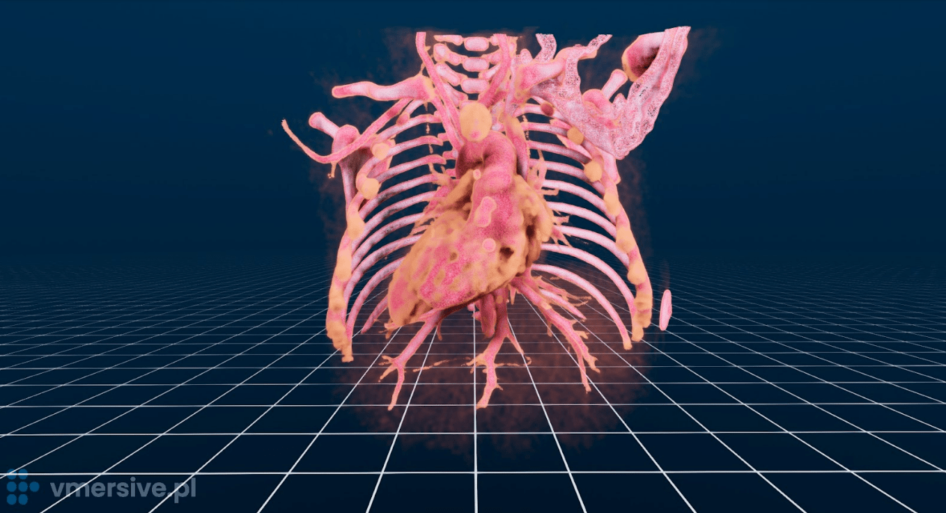
No Manual Segmentation

Automatic 3D reconstruction of any CT, MR or 3D Echo study in DICOM format. Instantaneous start of the simulation.

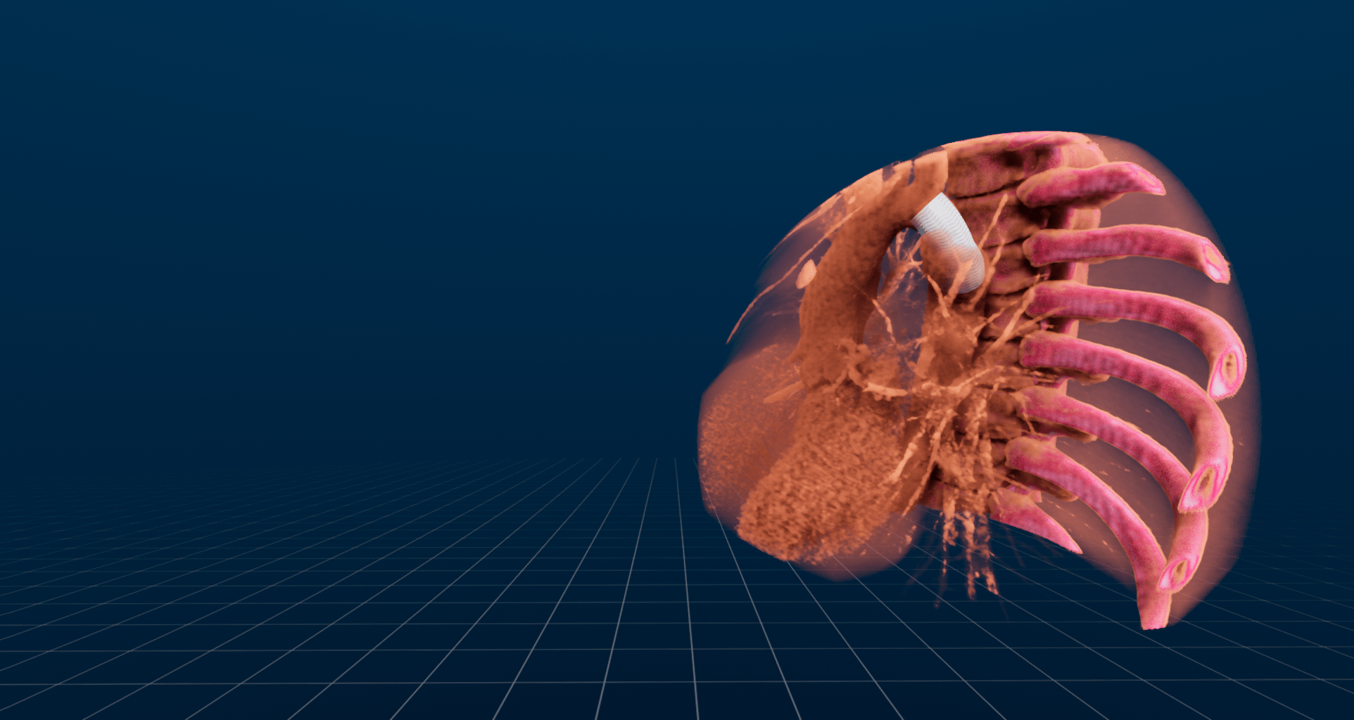
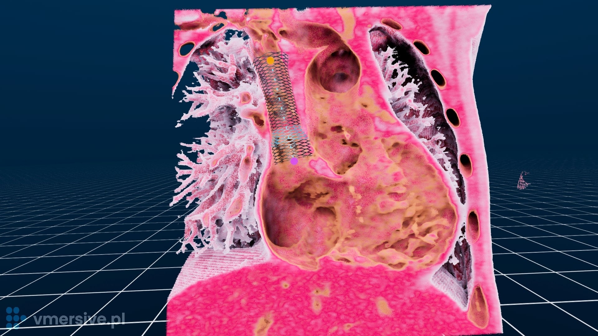
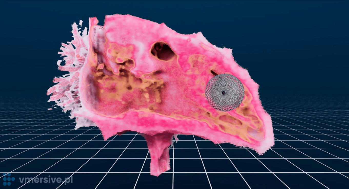
Library of Medical Devices

Integrated sizing sheets from major manufactures: stents with foreshortening tables, valves, VSD occluders, conduits.

Advanced Measurments

Accurate length of a line, curve, contour or value of an angle. Volume of any 3D region e.g heart ventricle.

Dynamic Studies

Full support of functional and multi-phase 4D studies to improve the realism of patient anatomy images.

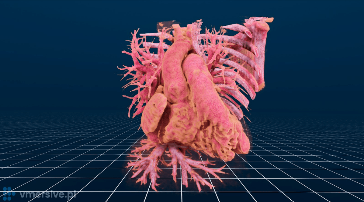
Full Anatomical Context

One click to observe heart interior and vessels, implanted devices, calcifications, airways or MPR.

Report Generation

Save the configuration of medical data, devices, measurements, screenshots and share it with the team.

Created Specifically For Doctors.

Previous VR Experience is not needed.

The process is simple: import DICOM files, wear a VR headset, and begin analyzing. Every interaction, such as slicing, cutting, measuring, or customizing a stent, has been carefully considered for ease of use.

In addition to the free trial we offer a free online training to help new users maximize their experience with the software.


Interventions & Surgeries Workflows

  • TPVI
  • CoA
  • SVD
  • DORV
  • VSD
  • Complex TGA
  • LVAD
Transcatheter pulmonary valve implantation
Analyze the Pulmonary Trunk in the full cardiac cycle.
Observe different anatomical contexts: vessels, hollow heart chambers, septal defects, calcified tissues
Evaluate optimal landing zone directly in 3D space
Simulate the angio view to find optimal RAO/LAO and CAUD/CRAN projections angles.
Coarctation of Aorta
Configure a stent with foreshortening table by selecting a specific model or design a custom one. Adjust stent’s parameters including flares.
Observe full cardiac cycle and interaction of the virtual stent with the entire context of anatomy including the airway system.
Simulate the angio view to find optimal RAO/LAO and CAUD/CRAN projections angles.
Sinus Venosus Defect
Customize a stent by either picking a pre-made model with a foreshortening table or creating your own design. Make necessary modifications to the stent’s size, shape, and flares.
Study the full cardiac cycle and examine how the virtual stent interacts with the entire anatomy, including the airway system.
Utilize simulation to determine the most favorable angles for RAO/LAO and CAUD/CRAN projections during the angio procedure.
Double outlet right ventricle (DORV)
Observe origins and connections of aorta and pulmonary and easily locate the VSD and determine its type.
Simulate different alternatives including the Blalock-Taussig shunt using conduits.
Verify simulated implants with full context anatomy including the airway system
Ventricular Septal Deffect
Analyze VSD in the full cardiac cycle.
Perform three dimensional measurements.
Select VSD occluder from Library of Medical devices
Simulate the angio view to find optimal RAO/LAO and CAUD/CRAN projections angles.
Complex Transposition Of The Great Arteries
Observe origins and connections of aorta, pulmonary trunk, pulmonary arteries and ductus arteriosus. Easily locate the VSD and its relative position to the great arteries.
Simulate different alternatives including: Blalock-Taussig shunt using conduits or Ductus arteriosus transcather stenting.
Verify simulated implants with full context anatomy including the airway system.
LVAD – HeartMate 3 implantation
Observe how the device fits within the heart anatomy, adjust the size of the ring.
Design the path of the outflow graft and driveline directly in 3D on a patient’s specific anatomy

Recommended by experts

Partners & Testimonials

Jenny Zablah Alabi, MD

Assistant Professor of Pediatrics, Interventional Cardiology,
Children’s Hospital Colorado, USA

“VMersive is a platform that allows quick and efficient procedural simulation with a user-friendly interface. Has multiple features that are specific for congenital heart disease with applications for surgical or percutaneous interventions..”

Michał Gałeczka, MD PhD

Paediatric cardiologist, research assistant,
Silesian Center for Heart Diseases, Zabrze, Poland

It is an outstanding modality of visualization and procedure simulation. Three-dimensional pictures in virtual reality give insight into specific and complex anatomy of Your patient. And it takes less than 15 minutes to get the proper pictures and to simulate the procedure.”


Trusted by:


Research Publications

Computer Modeling and Virtual Reality for Stent Development and Case Planning in Congenital Heart Disease
Gareth J Morgan, MD & Jenny Zablah, MD

Frequently Asked Questions

Product

Sales

IT / Legal

Reach out to us!


Direct Contact